Cutaneous Clues in Secondary Adrenal Insufficiency Associated with Mixed Connective Tissue Disease: A Case Report
Abstract
Secondary adrenal insufficiency (SAI) is a rare but potentially life-threatening complication of autoimmune connective tissue disorders such as mixed connective tissue disease (MCTD). We present a case of a middle-aged woman with MCTD who developed SAI. She exhibited cutaneous hyperpigmentation, particularly over the face, palmar creases, and soles, but lacked oral mucosal pigmentation. These findings, supported by biochemical tests and imaging, were consistent with a diagnosis of SAI rather than primary adrenal insufficiency (PAI). This case emphasizes the value of cutaneous findings in early recognition and differentiation of adrenal insufficiency subtypes.
Keywords
Secondary adrenal insufficiency, mixed connective tissue disease, hyperpigmentation, oral mucosa, ACTH, autoimmune
Introduction
Adrenal insufficiency may be classified as primary (PAI), secondary (SAI), or tertiary, depending on the level of the hypothalamic-pituitary-adrenal (HPA) axis affected. SAI results from deficient adrenocorticotropic hormone (ACTH) secretion by the pituitary and is often overshadowed by the clinical features of underlying autoimmune diseases like systemic lupus erythematosus (SLE), scleroderma, or MCTD [1]. Unlike PAI, SAI typically lacks features such as mucosal hyperpigmentation and hyperkalemia. Early recognition is critical to prevent adrenal crisis.
Case Presentation
A 40-year-old woman with a known diagnosis of mixed connective tissue disease (MCTD) presented with worsening fatigue, postural dizziness, and diffuse pigmentation over her face and extremities for the past three months. She denied gastrointestinal symptoms or significant weight loss.
Cutaneous Examination
-
Diffuse tan to bronze pigmentation over the forehead, malar areas, and perioral region (Image 1).
-
Hyperpigmentation of palmar creases a (Image 2).
-
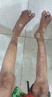
hypopigmented lesions over limbs.
-
Oral mucosa was spared; no pigmentation was noted on the buccal mucosa or gums.
-
Nails showed longitudinal ridging and mild dystrophy (Image 3).
Laboratory Findings
-
Morning cortisol: 4.1 µg/dL (low)
-
ACTH: 1.5 pg/mL (inappropriately low)
-
Electrolytes: low sodium and normal potassium
-
TSH: Within reference limits
-
Autoimmune panel: Positive ANA, anti-U1-RNP, consistent with MCTD
Imaging
MRI of the brain not done due to low resource settings and patient constrains. plan to obtain MRI Pituitary sequences.
Diagnosis
Based on clinical, biochemical, and imaging findings, a diagnosis of secondary adrenal insufficiency due to ?autoimmune pituitary involvement in MCTD was made.
Management
The patient was started on oral prednisolone (40 mg/day in divided doses) with education on stress dosing. She reported significant clinical improvement within 2 weeks of therapy initiation. Dermatologic changes gradually regressed over the subsequent 6 weeks. tolvaptan was tapered and plan to stop after 5 days.
Discussion
Hyperpigmentation in adrenal insufficiency results from increased ACTH stimulation of melanocortin-1 receptors in the skin. While this is a hallmark of PAI, similar pigmentation may occur in SAI due to prolonged hypocortisolism and overlapping autoimmune pathology [2]. However, oral mucosal pigmentation is considered more specific for PAI [3]. Our patient’s lack of mucosal pigmentation, together with inappropriately low ACTH levels, helped distinguish SAI from PAI.
Mixed connective tissue disease is known to have central nervous system manifestations, including pituitary involvement leading to hormonal deficiencies [4]. This case highlights the need for high clinical suspicion for HPA axis dysfunction in autoimmune diseases.
Conclusion
In patients with MCTD and nonspecific symptoms like fatigue and pigmentation, clinicians should maintain a high index of suspicion for secondary adrenal insufficiency. The absence of oral mucosal pigmentation, despite cutaneous changes, serves as an important clinical clue favoring SAI over PAI.


Nice case! Please share references, as I'm interested in seeing reference 2
ReplyDeleteIf TFTs were normal and clinical features of hypothyroidism were ruled out and she did not have any menstrual disturbances, then anterior pituitary may have been spared perhaps?
Was she on long-term steroid prior to this? The skins lesions look more Vitiligenous than SAI?
What drugs was she on for MCTD?
Why was prednisolone chosen?
Thanks for sharing.
Aditya