36M with decreased appetite, nausea and losse stools.
36 year old male farmer by occupation resident of xxxxx was brought to causality with cheif complaints of
1. Decreased appetite since 1 year
2. Nausea and vomitings since 6 months
3. Loose stools since 6 months
Since.
4. Tingling and numbness of both UL & LL since 10 days.
Patient was apparently alright 1 year back then initially he had decreased appatite associated with nausea which was gradually progresed to complete loss of appatite with in 5-6 months but able to still manage his daily routine activities. Then patient developed vomitings, 4-5 episodes per day , greenish , immediately after food intake. With food particles as contents. Not a/w blood in vomiting. Since then patient developed fear of eating.
Since 6 months patients had loose stools now increased in frequency since 2 months. 5-6 episodes per day, Watery consistency, large quantity, associated with greenish mucous with No blood in stools.
Last month with similar complaints patient went to one of the corporate centers in nearest city and evaluated further where he also had intra hospital hypoglycemic attacks.
Hb 9.6,
Alb 1 gm/dl,
sr. Cortisol 18.4
Ascitic tap was done :
AF - ADA 3.53, Sugars 81mg/dl, Gene expert Negative, proteins <2.0, cytology shows mixed inflammatory smear, negative for malignancy.
Usg abdomen : thickening of terminal ileal loop, moderate ascites.
UGIE : Hiatus Hernia, LAX LES, mild prolapse gastropathy with mild Duodenitis.
Colonoscopy : Non specific segmental colitis.

IgA- TTG 0.5
Fecal calprotectin 65.3
CECT Abd : Diffuse long segment circumferential wall enhancement of small bowel loops
MRI Abd : Howed subcentrimetric lymphadenopathy and tiny rt renal cortical cyst.
H/o significant weight loss upto 20kgs with in 1 year.
H/o blood in stools present once a while
H/o fever on and off.
Not a known case of DM, HTN, TB
H/O multiple admissions for B/l lower limb weakness ( ? Recurrent hypokalemia ) records not available.
Personal history :
Appetite : decreased
Diet : mixed
Sleep : inadequate
Bladder : normal UO
Bowel movements: chronic diarrhoea
Addictions : occasional alcoholic and smoker.
General examination:
Pallor: present
Icterus: absent
Cyanosis : absent
Clubbing : preeent
Lymphadenopathy : absent
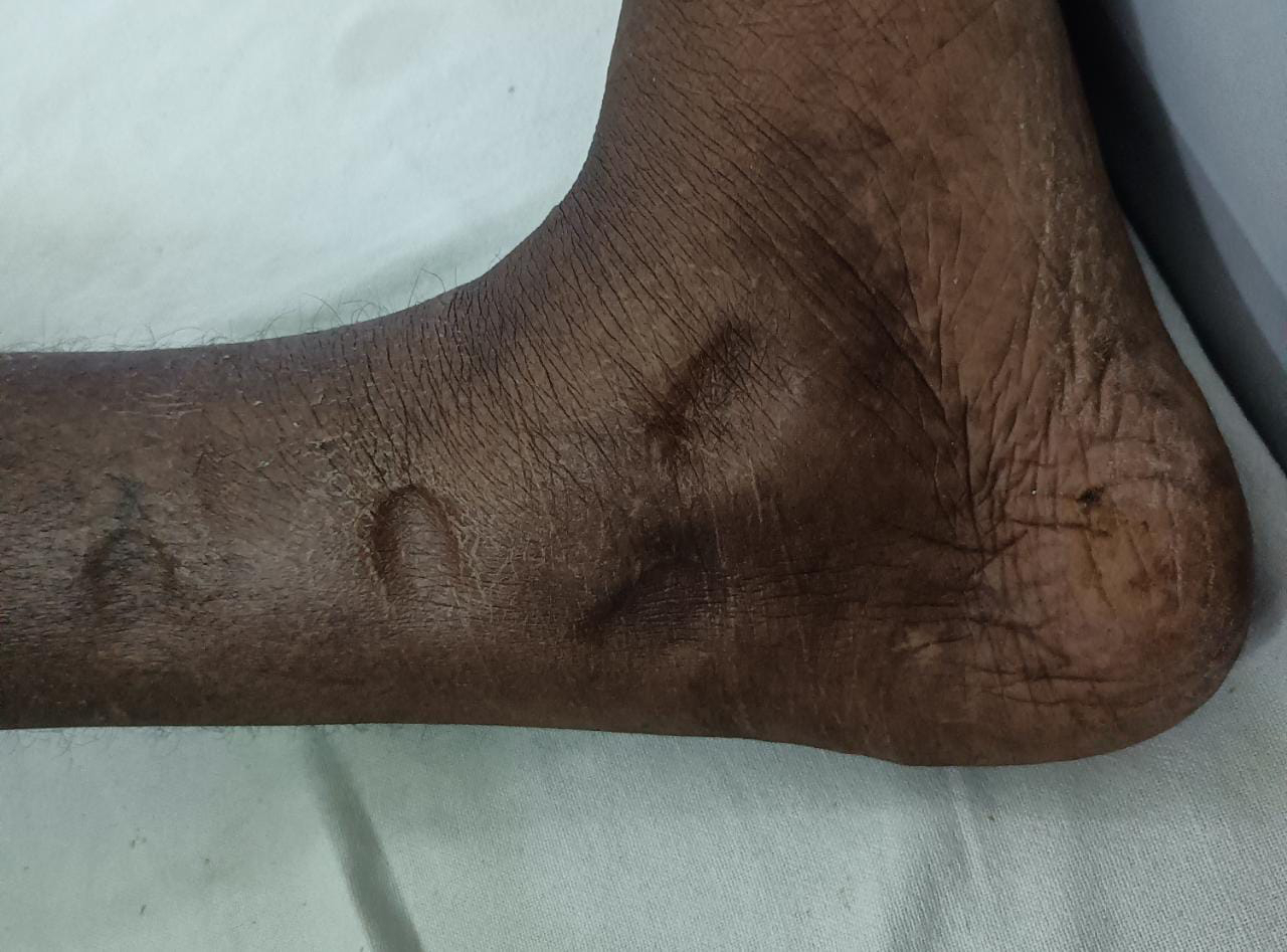
Edema : B/L pedal edema present upto knee

Vitals:
Temperature: afebrile
Pulse: 110bpm
Blood pressure: 90/60 mm of hg
Respiratory rate : 20 bpm
SpO2 : 98 on RA
GRBS : 85 MG/DL
Systemic examination:
Cardiovascular system
JVP - not raised
Visible pulsations: absent
Apical impulse : left 5th intercostal space in midclavicular line.
Thrills -absent
S1, S2 - heart sounds heard
Pericardial rub - absent


Respiratory system:
Patient examined in sitting position
Inspection:-
oral cavity- Normal ,nose- normal ,pharynx-normal
Shape of chest - normal
Chest movements : bilaterally symmetrically reduced
Trachea is central in position.
Palpation:-
All inspiratory findings are confirmed
Trachea central in position
Apical impulse in left 5th ICS,
Chest movements bilaterally symmetrical
AUSCULTATION
BAE+, NVBS

Abdomen examination:
INSPECTION
Shape : mild distended
Umbilicus:normal
Movements :normal
Visible pulsations :absent
Skin or surface of the abdomen : normal
PERCUSSION- tympanic
AUSCULTATION :bowel sounds heard
Usg abdmen :

Labs :

ABG :
pH 7.57
pCO2 : 19.8
PO2 : 114
Hco3 : 18.5
St. Hco3 : 22.8
Spo2 : 98
Stool for occult blood : positive.
Diagnosis :
Chronic diarrhoea under evaluation.
Treatment :
Iv fluids 1 unit NS @ 75ML/HR
Inj. Zofer 4 mg sos
Inj. PAN 40 mg IV BD
Inj. Optineuron 1 amp in 100ml NS IV OD
Inj. PCM 1 gm IV sos if Temp >102F
Tab. Loperamide 4mg po BD
Tab. Sporolac DS po TID
Ors sachets 1 packet in 1 ltr water.
HIGH Protein Deit.
GRBS 4th hourly.


Outside hospital colonoscopy was done biopsy report awaited.

11/07/22.
Stool microscopy reveals eggs of Capillaria Philippinesis.


Discussion
[7/11, 12:47 PM] saicharankulakarni:
Human intestinal capillariasis caused by Capillaria philippinensis is an endemic disease in Philippines[1] and Thailand.[2] C. philippinensis infection is mainly acquired by ingestion of raw, undercooked, small fresh water or brackish water fi sh, in which larval forms of the parasite develop. This infection mainly involves small intestine (jejunum) and patients suffer from chronic diarrhoea, protein losing enteropathy, borborygmus and electrolyte loss.[1] If early diagnosis and treatment is not given, it can be fatal.
Only two cases have been reported from India. No case was reported from Andhra Pradesh. The fi rst case from India was reported by Kang et al. in 1994 from Vellore[6] and the second case by Rana et al. in 2009 from Chandigarh.[7] This is the third case report from India and the fi rst case from Andhra Pradesh to the best of our knowledge
[7/11, 12:48 PM] saicharankulakarni:
C. philippinensis is a nematode belonging to class Adenophorea, subclass Enoplia, order Trichurida and family Trichinellidae.[8] In the life cycle of C. philippinensis, fi sh eating birds are natural defi nite hosts. Adult worms present in the intestine of birds release ova. Bird droppings along the fl yways disperse these eggs into water bodies, where f i sh become infected. Larval forms develop in fi sh and it is the source of infection to man and bird.
C. philippinensis infection is mainly acquired by ingestion of raw, undercooked, small fresh water or brackish water fish.
Simple wet mount examination of stool sample and identifi cation of ova, larvae and adult worm in the stool sample can clinch the diagnosis. But ova of C. philippinensis need to be differentiated from those of Trichuris trichura. Ova of C. philippinensis can be identifi ed by their peanut shape, fl attened mucous plugs and striations in the wall. Adult worms vary in sizes from 2 to 5 mm. Male worms are shorter (1.5 – 3.9 mm) compared to female worms (2.3 – 5.3 mm). They are identifi ed by their characteristic stichosome, a muscular oesophagus surrounded by rows of stichocytes. Male worms have single sheathed spicule. Female uterus contains numerous thick-shelled eggs and thin-shelled eggs with or without embryos or larvae. Larvae found in stool sample in different stages of development and hence are diffi cult to identify as C. philippinensis larvae.[1]
Patient was continued on T. Albendazole with electrolytes replacement. On day 2 after starting albendazole patient dryness of mouth decreased but appatite still didn't improved .
20/7/22
http://drkulkarnimd.blogspot.com/2022/07/36m-with-decreased-appetite-nausea-and.html
S - loose stools subsided, appatite improved, generalised weakness present.
O - vitals unremarkable,
Na 129 K 2.6 cl 103 ionised ca 0.74.
A - chronic diarrhoea 2to C.philipiensis( resolved ) with dyselectrolytemia.
P - mobilising patient. To send Sr. Albumin and total proteins.


Comments
Post a Comment