35 F with pain abdomen
35 year old with pain abdomen
CHIEF COMPLAINTS :
35 year old female who is a wig maker and goes to part
time work in paddy and cotton field’s presented to the Emergency department
with acute history of pain abdomen & vomiting’s since 4 days
INTRODUCTION
INTO BIOPSYCHOSOCIAL ASPECTS OF THE CASE:
Activities of
daily leaving before illness:
Patient was apparently asymptomatic 7 years (2016) back where
here daily routine before illness was waking up at 4 am after cleaning house
and fresh-up herself, prepare food for the family. She used to have IDLI, DOSA,
RAAGI JAVA as her breakfast and take rest for few minutes and enjoys working
for family household needs like cooking, laundry, cleaning home etc. she
prepare dinner and get to sleep by 10pm.
History of presenting illness:
Down the line 7 years back in 2016 while she was going in
auto she was having conversation with driver (cousin) and met with an road
traffic accident where auto was hit by lorry from back. She had sustained
injury to her head with minor superficial injuries on face, fear of impending
doom ( first psychological impact ) with no loss of consciousness, no history
of headache , no history of vomiting’s, then she was shifted to nearest medical
care and was incidentally diagnosed with type -2 Diabetes mellitus.
After an year in 2017 her husband developed severe
headache for 3 months and attaining medical check-up she was counseled that her
husband had some lesions in brain (described as blood clots) and he will not
survive for more than 3-6 months (with no motor deficits) and he passed away
after 2 months (second psychological impact).
Following demise of her husband her life was taken
another path where here daily routine was scheduled to earn for living (third
psychological impact with associated social factors).
Impaired
activities after death of her husband:
Wakes up around 4am in the morning, gets ready for work
with frequent skipping of breakfast compared to past. She starts her work at
8-9am sell ornaments, sometimes work in paddy fields (for planting and other
farming work). She get back to home by 11:00 am have some snacks and look after
her elder daughter while she was pregnant and younger daughter who is pregnant
now. Household work, and sits at a retail shop or play with her grand-daughter.
She bags around 4000-6000 per month with net savings of 500-700 rps/- per week
approximately.
She used to raise money 10000-20000 based on need at
local groups for her daughters health check-ups, sons college fees and or daily
household needs which was cleared by her savings.
She developed frequent burning sensation in her chest
localized to epigastric region associated with bloating, belching and altered
bowel habits (constipation with once in 2 days since last few months)
Possible
stressors and outcomes:
Impaired daily activities
Physical and emotional stress
Social stress (financial burden)
Altered food and bowel habits
Dyspepsia and frequent use of antacids.
Acute History
of presenting illness:
Now, presenting with complaints of pain abdomen originated
at left hypochondrium and lumbar region (VAS 7 score) then became diffuse in
type over 10 to 12 hours (VAS 10
Worst pain she ever had), squeezing type of pain with
occasional coliky type, associated with vomiting’s 2-3 episodes non bilious ,
non-projectile, non-blood tinged with food particles as content. Passing stools
small amount (as fecal pellets of goat - described by patient) didn’t pass
flatus since 2 days.
history of bloating, belching since 5 years using
antacids.
Passage of hard stools since 2 months (Bristol stool
chart - type 1)
MARRIAGE
& OBSTETRIC HISTORY:
Married at the age of 17 years , non-consanguineous.
FIRST child at 18 years - death of the first child at
24years due to varicella zoster.
2nd child at age of 21 years - gave birth to
female child , now married
3rd child at the age of 23 years -gave birth to female
child , now married
4th child at the age of 24years - gave birth to male
child currently 10th class.
PERSONAL
HISTORY:
Mixed diet, appetite reduced
Constipation since 5 months
NON SMOKER AND NON ALCOHOLIC
FAMILY
HISTORY:
Mother is hypertensive and expired due to cerebrovascular
accident and post stroke complications after 6 months.
DRUG HISTORY:
weekly she used to take 2-3 sachets of ENO (antacids)
AT
PRESENTATION:
Patient is conscious, coherent and Co-operative well
oriented with time, place and person.
VITALS AT
PRESENTATION:
Febrile to touch 99.1 F
Blood pressure: 110/80mmhg in right upper arm supine
position.
Pulse rate: 119 per minute, regular, normal volume
Respiratory rate: 24 cycles per minute (pain induced
tachycardia)
Room air saturation: 92%
GENERAL
EXAMINATION:
Looks grossly dehydrated with sunken eyes, dry oral
mucosa and tongue with delayed capillary refill time (more than 3 seconds)


Pallor present
bilateral pitting type of pedal Edema present with
scratch marks on both lower limbs (pruritus due to possible diabetic
dermopathy)
No icterus, cyanosis, clubbing, lymphadenopathy.
She used to colour her hair since 8 years (early
whitening of hairs at 28 years – probable zinc and other nutritional
deficiencies)
SYSTEMIC
EXAMINATION:
EXAMINATION OF THE ORAL CAVITY
NO Oral thrush, NO tonsilar enlargement &
pharyngeal deposits, NO post nasal drip, NO fetor hepaticus, Fair oral hygiene,
no dental caries and no gum hypertrophy.
ABDOMEN:


INSPECTION:
1. Shape – distended-uniform
2. Flanks – full
3. Umbilicus – central, Shape-slit like and
nodules.
4. Skin – stretched, no scars & sinuses,
striae present, scratch marks.
5. No Dilated veins – front/back
7. Movements of the abdominal wall,NO visible
gastric & intestinal peristalsis.
8. Hernial Orifices - normal
9. NO Renal angle tenderness

PALPATION:
Superficial Palpation – Tenderness present at
left hypochondrium and lumbar region, no local rise in temperature.
Deep Palpation
1.
Liver: inferior edge palpable, smooth non nodular.
2.
Spleen non
palpable and non-tender when palpated in the Left Hypochondrium.
3.
Kidney
non-tender and non palpable in the Right/Left Lumbar.
4. Abdominal Girth - 79cms
5.
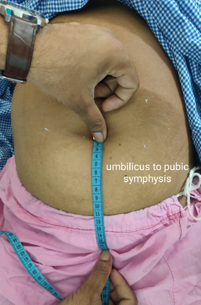
Distance
between the Xiphisternum-Umbilicus and Umbilicus-Pubic Symphysis equal 17 cms.

6.
No Murphy’s
Punch/Renal angle tenderness.
PERCUSSION:
1. Puddle’s sign present suggestive of mild
fluid collection in abdomen.
2. Percussion of Liver for Liver Span - 13cms
AUSCULTATION:
1. Bowel sounds – sluggish
2. No Bruit – Aortic, Hepatic, Renal Bruit
3. No Venous Hum.

OTHER
SYSTEMS EXAMINATION:
CARDIOVASCULAR SYSTEM:
No raised Jugular venous pressure
Apex beat in 5th Intercostal space on left mid clavicular
line
S1 and s2 heard. NO murmurs.

EXAMINATION OF RESPIRATORY SYSTEM:
No tracheal deviation
Respiratory movements are bilateral symmetrical
Resonant percussion in all lung field’s
Normal vesicular breath sounds.

EXAMINATION OF NERVOUS SYSTEM:
Higher mental functions intact
All Cranial nerves on both sides are intact
No motor deficits
Sensory system :
Fine touch - absent below both ankles
Vibration - delayed in both lower limbs ( 6sec at ankle, 8 sec at knee ) and upper limbs ( 9sec in upper limb )
Gait normal
PROBLEM STATEMENT:
A 35 year old diabetic female with acute history of
diffuse pain abdomen with nausea followed by vomiting and peripheral neuropathy.
Differentials:
1. Acute
intestinal pseudo-obstruction (ogilvie syndrome)
2. Gastroesophageal
reflux disease (GERD/?LAX lower esophageal sphincter)
3. Possible Acute pancreatitis (extends from hilum of spleen to epigastrium)
4. Peripheral neuropathy secondary to Diabetes
Gastroenterologist consultation was taken :
Advised for IgG4 antibodies , ANA and MRCP.
Investigations :


Acute colonic pseudo obstruction ( Ogileve syndrome - resolved )
Gastroesophageal reflux disease


Comments
Post a Comment